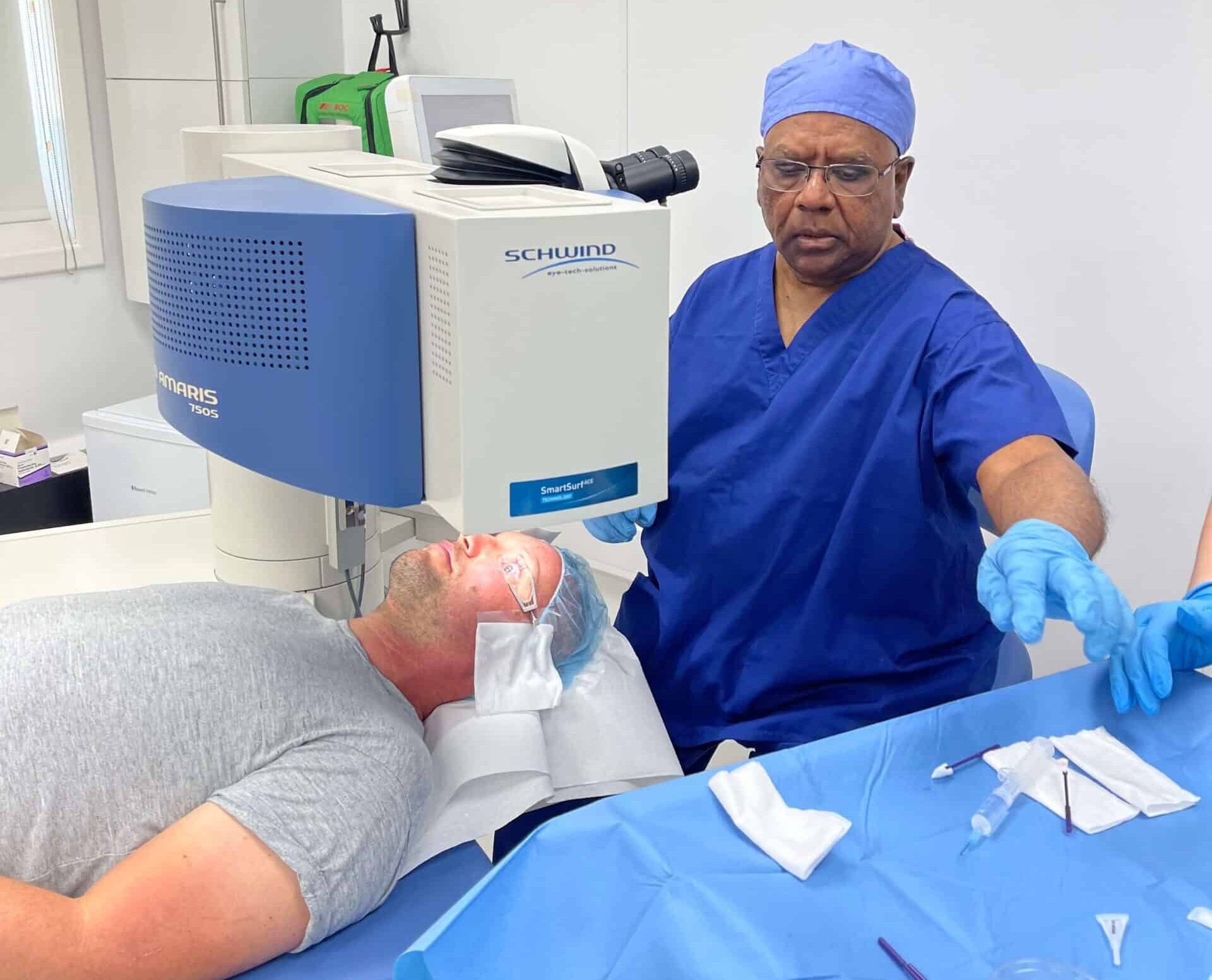
Myths about keratoconus and cross-linking often overshadow the facts about this significant eye condition that affects 1 in 500 people. Many misconceptions exist about the condition and its treatment options, even though it’s relatively common. Making informed decisions about your eye health requires you to understand the reality behind these myths, especially if you have a keratoconus diagnosis or are looking into treatment options.
Most patients who undergo corneal cross-linking experience stabilisation or improvement in their corneal shape and visual function. This highly effective treatment can lead to better visual acuity and might reduce the need for corrective lenses. People of all ages can develop keratoconus, though it usually starts between ages 10 and 25. This makes accurate information about cross-linking treatment vital for everyone, no matter when symptoms first appear.
This piece separates medical facts from fiction about keratoconus and cross-linking treatment. You’ll learn what this procedure can achieve, understand what happens during cross-linking, and discover why treating it early matters. Expert care and proper understanding make it possible to manage this condition effectively.

Understanding Keratoconus and Cross-Linking
Your eye’s cornea works as a clear window at the front, naturally keeping a round dome shape. If you have keratoconus, this delicate structure gets thinner and bulges outward into an irregular cone shape. This condition starts during puberty or early adulthood without inflammation. It usually gets worse until the mid-30s before it stabilises on its own.
What is keratoconus?
Keratoconus happens when collagen fibres that keep your cornea’s strength and shape become weak. The cornea thins out and pushes forward, which disturbs your vision. The original signs might include blurry vision and worsening short-sightedness with irregular astigmatism. It also causes fine vertical lines in the cornea called Vogt striae and leaves an iron deposit ring known as a Fleischer ring. European rates of keratoconus range between 5 and 23 per 100,000 people each year. Dutch studies show about 265 cases per 100,000 people. The condition links to several body-wide disorders including atopy, Down syndrome, and Marfan syndrome.
How does corneal cross-linking work?
Corneal cross-linking (CXL) stands out as a breakthrough treatment. It’s the only proven way to stop keratoconus from getting worse. The treatment uses vitamin B2 (riboflavin) eye drops on the cornea and controlled ultraviolet-A (UVA) light exposure.

This mix creates a chemical reaction that builds new bonds between collagen fibres, linking them together. These extra bonds make your corneal structure stronger and stop further thinning and bulging. Doctors typically use 30 minutes of UVA exposure at 3mW/cm² intensity, which delivers 5.4 J/cm² of total energy. CXL shows remarkable results. Studies prove it stops progression in more than 90% of treated eyes. Some patients’ corneal shape actually improves.
Why early diagnosis matters
Finding keratoconus early makes a big difference because CXL works best before your cornea suffers serious damage. Catching the condition at its start lets doctors step in quickly to prevent permanent vision loss. Today’s diagnostic tools like corneal topography can spot tiny corneal changes. They find keratoconus before you notice any symptoms. Scheimpflug tomography offers detailed 3D corneal views. It measures thickness and checks biomechanical properties with great accuracy. Quick treatment helps you avoid problems like corneal scarring that make vision correction with glasses or contacts harder. Getting help early substantially cuts your chances of needing a corneal transplant. Without treatment, about 20% of progressive cases need this procedure.
4 Common Myths About Keratoconus & Cross-Linking
People often misunderstand keratoconus and cross-linking beyond their simple basics. Let’s get into the most common myths that make patients worry without reason.
Myth 1: Cross-linking is painful and risky
Patients worry about pain during corneal cross-linking. The procedure doesn’t hurt because anaesthetic eye drops completely numb your eye. You might feel some discomfort after treatment as the surface layer heals, but this lasts only 24-48 hours. The safety record speaks for itself – complications happen in less than 3% of cases. More than 90% of treatments succeed, which makes this procedure both safe and effective for most patients.
Myth 2: It only works in early or mild cases
Cross-linking can work even in advanced stages of keratoconus, unlike what most people believe. Studies of patients with advanced progressive keratoconus (corneas with Kmax≥58 D) show that cross-linking stabilised the condition in 96.6% of eyes. The data shows that accelerated cross-linking has higher success rates in eyes with steeper preoperative measurements, showing its effectiveness in cases of all stages.
Myth 3: Contact lenses are enough to stop progression
This myth about keratoconus management comes up most often. Scleral lenses and rigid gas permeable lenses improve vision quality by a lot, but they can’t stop the disease from getting worse. Some evidence suggests flat-fitting corneal lenses might damage an ectatic cornea. Corneal cross-linking remains the only treatment that tackles the mechanisms of keratoconus by making the corneal structure stronger.
Myth 4: Cross-linking cures keratoconus
Cross-linking doesn’t cure keratoconus. The treatment stabilises the cornea and prevents further damage. The goal focuses on stopping progression rather than reversing existing changes or restoring vision. About 10% of patients might need another procedure to fully stop progression. Most patients still need glasses or contact lenses for the best vision after treatment.
What Cross-Linking Can and Cannot Do
Your CXL treatment will work better when you know what to expect from it. This powerful procedure has specific benefits and limitations you should know about.
Slows or halts progression, not a cure
CXL works to stabilise keratoconus instead of reversing existing damage. Studies show CXL stops progression in more than 90% of treated eyes. These numbers show a soaring win, but 7-8% of patients might still see their condition progress after treatment. CXL doesn’t eliminate keratoconus – it makes your cornea stronger to stop further damage.
May improve vision but not always
Vision improvement isn’t the main goal, but about 45% of patients see better corneal shape after CXL. Research shows vision gains between one and two Snellen lines within 1-4 years after treatment. These improvements happen slowly and become noticeable about three months after your procedure. You’ll need patience since the full effects take 6-12 months to show up.
Does not eliminate the need for glasses or lenses
You’ll definitely need glasses or contact lenses after CXL. The treatment stabilises your cornea but doesn’t fix existing vision problems. Your prescription might change after treatment, but you’ll still need corrective eyewear to see well. Think of CXL as a way to preserve your current vision rather than a fix for your need for vision correction.
Repeat procedures may be needed in rare cases
The largest longitudinal study shows retreatment rates of 8-14% over 15 years. A second round of cross-linking can help stabilise keratoconus that keeps progressing after the original treatment. Regular check-ups are vital – first every six months, then yearly – to spot any signs of renewed progression.
Why Choose Precision Vision London for Cross-Linking
Expertise and quality of care matter the most to patients who need corneal cross-linking treatment. Precision Vision London emerges as a premier choice that delivers effective treatment to keratoconus patients.

Expert surgeons with years of experience
Dr. CT Pillai leads Precision Vision London with over 30 years of ophthalmic expertise. He stands among a select group of UK surgeons who hold dual fellowships in corneal and refractive surgery. Since 2007, Dr. Pillai has mastered the art of collagen cross-linking treatment. His innovative achievements include performing bilateral laser eye surgery as one of the country’s first surgeons.
Advanced diagnostic and treatment technology
The clinic uses state-of-the-art equipment for diagnosis and treatment. Advanced diagnostic tools like corneal topography and optical coherence tomography (OCT) create precise maps of your cornea’s shape. These tools help detect keratoconus early and monitor its progression accurately. Patients experience superior outcomes from cross-linking procedures thanks to this technological edge.
Personalised care plans for every patient
Quality takes precedence over quantity at Precision Vision London, and each patient receives individualised care. Your treatment starts with a detailed consultation to check suitability and create a personalised treatment plan. This customised approach continues through aftercare. Patients can access the country’s largest independent optometrist network.
Trusted by patients across the UK
Patient expectations are exceeded by the clinic’s dedication to excellence. Dr. Pillai maintains an outstanding 4.98 out of 5 stars rating from 154 patient reviews. Patients often report major improvements in their vision and exceptional support throughout their treatment.
Conclusion
Making informed decisions about your eye health starts with understanding keratoconus and cross-linking treatment. Medical facts need to be separated from fiction when you think over treatment options. Keratoconus can feel overwhelming initially, but early intervention through cross-linking will halt progression in over 90% of cases.
Cross-linking can’t reverse existing damage completely. However, it stabilises the cornea and stops further deterioration. Many patients see improvements in their corneal shape and visual acuity after the procedure. Without doubt, this has made cross-linking the gold standard treatment for progressive keratoconus in the UK.
Your regular eye examinations play a vital role, especially when you have risk factors or a family history of the condition. Treatment outcomes and vision quality improve substantially with early diagnosis. Eye assessments should be your priority, particularly if you notice changes in your vision.
Dr. CT Pillai at Precision Vision London brings over 30 years of specialised experience to keratoconus treatment. Advanced diagnostic equipment at the clinic gives accurate assessment of your corneal condition. Their personalised care approach creates treatment plans that address your specific needs. The clinic’s exceptional patient satisfaction ratings show the quality of care you’ll receive throughout your treatment experience.
Note that keratoconus needs ongoing management, but proper treatment can stabilise your condition and maintain visual function. Expert care and advanced technology at Precision Vision London help you face keratoconus confidently. Your eye health stays in capable hands. Evidence-based treatment offers the best path forward for anyone diagnosed with this common yet manageable condition.
Key Takeaways
Understanding the facts about keratoconus and cross-linking treatment helps you make informed decisions about this manageable eye condition that affects 1 in 500 people.
- Cross-linking successfully halts keratoconus progression in over 90% of cases, making it the gold standard treatment for this condition
- The procedure strengthens corneal structure but doesn’t cure keratoconus—most patients still need glasses or contact lenses afterwards
- Early diagnosis is crucial as cross-linking works best before significant corneal damage occurs, potentially avoiding corneal transplants
- Cross-linking is safe with complications in less than 3% of cases and can be effective even in advanced keratoconus stages
- Contact lenses alone cannot stop disease progression—only cross-linking addresses the underlying corneal weakening that causes keratoconus
With proper expert care and realistic expectations, keratoconus can be effectively managed through cross-linking treatment, preserving your vision and preventing further deterioration of this progressive condition.
FAQs
Q1. How effective is corneal cross-linking in treating keratoconus? Corneal cross-linking is highly effective, halting keratoconus progression in over 90% of treated eyes. It strengthens the corneal structure by creating new bonds between collagen fibres, effectively stabilising the condition.
Q2. Can cross-linking improve vision in keratoconus patients? While not the primary goal, approximately 45% of patients experience improved corneal shape after cross-linking. Some may notice vision gains within 1-4 years post-treatment, but these improvements typically develop gradually.
Q3. Is corneal cross-linking a painful or risky procedure? The procedure itself is generally pain-free due to anaesthetic eye drops. Some discomfort may occur for 24-48 hours afterwards as the eye heals. Cross-linking has an excellent safety profile, with complications occurring in less than 3% of cases.
Q4. Can keratoconus be treated at any stage with cross-linking? Contrary to popular belief, cross-linking can be effective even in advanced stages of keratoconus. However, early intervention is ideal as it can prevent significant corneal damage and potentially avoid the need for corneal transplants.
Q5. Will I still need glasses or contact lenses after cross-linking? Most patients still require glasses or contact lenses after cross-linking. The treatment stabilises the cornea and may improve its shape, but it doesn’t necessarily correct existing refractive errors. Your prescription might need adjustment after treatment.
Authors & Reviewer
-
 Olivia: Author
Hi, I'm Olivia, a passionate writer specialising in eye care, vision health, and the latest advancements in optometry. I strive to craft informative and engaging articles that help readers make informed decisions about their eye health. With a keen eye for detail and a commitment to delivering accurate, research-backed content, I aim to educate and inspire through every piece I write.
-
 Dr. CT Pillai: Reviewer
Dr. CT Pillai is a globally recognised ophthalmologist with over 30 years of experience, specialising in refractive surgery and general ophthalmology. Renowned for performing over 50,000 successful laser procedures.

