
Keratoconus Treatment in London
Keratoconus is a known, manageable eye condition, and treatment can usually stop it from progressing. If you have recently been diagnosed, our keratoconus treatment in London starts with a detailed assessment to understand how the cornea is changing and whether treatment is needed.

- Comprehensive consultation & corneal mapping
- 12-month aftercare programme included
- 24-hour post-treatment telephone access







For keratoconus that is continuing to progress, corneal cross-linking (CXL) strengthens the cornea to help slow or halt further progression. Your care is led by a Consultant Ophthalmic Surgeon at our Harley Street clinic, who will explain what your scans show and whether treatment is appropriate for you.
What is keratoconus?
The cornea is the clear, curved surface at the front of your eye that helps focus light. With keratoconus, the cornea gradually becomes thinner and steeper, which can give it a more cone-like shape. As the shape changes, light does not focus as evenly and vision can become blurred or distorted.
Keratoconus commonly develops during the teenage years or in your twenties. Being diagnosed when you are young is therefore not unusual. The condition behaves differently from person to person, and some people's corneas change more than others. For some people keratoconus stabilises over time, while for others, if cross-linking is not undertaken, the condition can progress to the point where a corneal graft (corneal transplant) is required.
The exact causes of keratoconus are not fully understood. Genetics may play a part, while frequent eye rubbing has also been associated with the condition and its progression.

Reasons people come to us with keratoconus symptoms
Keratoconus is sometimes discovered during a routine eye test before someone has realised anything unusual is happening. At other times, changes in vision lead people to have their eyes checked.
Common reasons people come to us include:
- Distorted vision
- Increasing short-sightedness or astigmatism
- Frequent changes to a glasses or contact lens prescription
- Greater sensitivity to bright light or glare
Having one of these symptoms does not tell us on its own whether keratoconus is progressing. Detailed measurements of the cornea are used to understand what is happening in each eye.
How is keratoconus diagnosed?
Your assessment includes an eye examination and corneal mapping. These scans create a detailed picture of the shape and thickness of your cornea, allowing the doctor to look for changes associated with keratoconus.
The scans are straightforward and take only a short time. Previous measurements can also be compared with new scans to see whether the shape of the cornea has changed over time.
Once the assessment is complete, your doctor will explain what the results mean. If your keratoconus appears stable, monitoring may be appropriate. If the scans show that it is progressing, corneal cross-linking may be discussed.
Corneal cross-linking treatment for keratoconus
Corneal cross-linking, usually shortened to CXL, is the main treatment used to slow or halt progressive keratoconus. You may also see it referred to as cross-linking surgery.
Book Your Assessment
CXL uses riboflavin (vitamin B2) drops together with controlled ultraviolet light. These work together to create stronger links between collagen fibres within the cornea, increasing its strength and helping it retain its shape.
The purpose of CXL is to slow or halt the progression of keratoconus.
CXL is not carried out with the expectation of improving eyesight. Some people notice changes in their vision after treatment, although this cannot be predicted and is not the purpose of the procedure. Glasses or contact lenses will still be needed afterwards.
There are two approaches to cross-linking: epithelium-on (epi-on) and epithelium-off (epi-off). Epi-off is the more effective of the two, with a higher success rate of halting progression with a single procedure, which is why it is the approach of choice at Precision Vision. This means the thin surface layer of cells on the cornea is gently removed before riboflavin is applied, allowing it to enter the corneal tissue before the UV stage of treatment.

Who is CXL treatment suitable for?
CXL is generally considered when keratoconus is progressing rather than at early stages. Corneal scans are important because they allow the doctor to look for changes in shape and thickness rather than recommending treatment simply because keratoconus has been diagnosed.
Age also forms part of the assessment because keratoconus commonly develops when the cornea is younger and more flexible. Some people have keratoconus that remains stable and may only need regular monitoring.
At Precision Vision, collagen cross-linking is offered to patients aged 18 and over whose keratoconus has not reached an advanced stage. Your doctor will review your individual measurements and explain whether CXL is appropriate, and suitability is always confirmed by the surgeon after a full assessment at your consultation.
Other keratoconus treatments
CXL is used to address progression, while other treatments can help manage the effect keratoconus has on vision or the shape of the cornea.
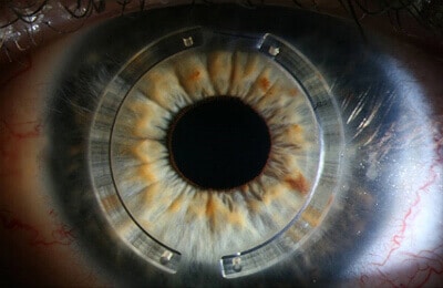
Options at Precision Vision include specialist contact lenses such as Kerasoft lenses, corneal implants (INTACS and Kerarings), and treatments to correct a residual prescription: implantable contact lenses (ICL), typically for patients under 40, and refractive lens exchange (RLE), typically for those aged 45 and over.
Specialist contact lenses can help provide clearer vision by correcting some of the visual effects caused by an irregular cornea. Corneal implants are small inserts placed within the cornea that apply gentle pressure to reshape the bulge into a more regular shape. This can make contact lens wear comfortable again, and for some patients it means returning to soft toric lenses rather than needing hard or rigid gas permeable (RGP) lenses. There can be a slight improvement in vision, although this is not the purpose of the procedure. Corneal implants may be considered for some patients once keratoconus is stable.
Your doctor will explain what each option is designed to achieve and whether it is relevant to your eyes. These treatments have a different purpose from CXL, which is used to support the cornea and slow or halt progression.
What happens on the day of CXL treatment?
Knowing what will happen before you arrive can make the treatment easier to prepare for. If both of your eyes need cross-linking, each eye is treated individually. Your team will explain the treatment plan beforehand, and there will be opportunities to ask questions.
Before treatment begins
Anaesthetic eye drops are used to numb the surface of the eye. An eyelid holder is then used to help keep the eye open, so you do not need to worry about accidentally blinking during treatment.
At the Precision Vision eye clinic, CXL currently takes around 60 minutes per eye. The exact time can vary depending on the individual treatment.
During the procedure
With the eye numb, the thin surface layer of the cornea is gently removed. Riboflavin drops are then placed onto the eye so they can be absorbed into the cornea.
The eye is exposed to a controlled amount of ultraviolet light, with additional drops used as needed during this stage.
You will be awake during the treatment. The anaesthetic drops are used to keep the surface of the eye numb, although people experience sensations differently. The clinical team will stay with you and explain what is happening as the procedure progresses.
Once treatment is finished, a soft bandage contact lens is placed over the cornea. This protects the surface while it begins to heal.
What does CXL feel like afterwards?
Once the anaesthetic drops wear off, the treated eye can feel sore or gritty. It may also water more than usual and become sensitive to light. Discomfort is often most noticeable during the first few days, although experiences vary between patients.
You will be given instructions about your prescribed eye drops and pain relief before you go home. The bandage contact lens remains in place while the surface of the cornea heals and is then removed by the surgeon at your first follow-up appointment.
The team will explain what to expect during the first few days before you leave the clinic.
When will my vision return to normal?
Vision is usually hazy at first. This can happen during the early healing period and does not mean the treatment has failed.
Your sight should gradually become clearer, although vision can fluctuate for several weeks. Your glasses prescription may continue to change while the cornea settles, sometimes for several months.
Your doctor will tell you when it is appropriate to have your prescription checked again or return to contact lens wear.
When can I go back to work or study?
It is sensible to allow around a week away from work after CXL. Students may also need some time away from college or university, depending on how comfortable the eye feels.
Reading and using screens can feel tiring during the early recovery period because vision may still be blurred or sensitive to light. You can gradually return to your usual routine as the eye becomes more comfortable.
Your team will give you individual guidance about returning to exercise and driving. Advice can vary depending on how your eye is healing.
Risks and considerations of treating keratoconus
CXL is a well-established treatment for progressive keratoconus, although every medical procedure has potential risks. Your doctor will discuss these before treatment so you can make an informed decision.
During the first few days, soreness and light sensitivity are expected while the surface of the cornea heals. Vision can also be blurred or variable during recovery.
Less common complications can include:
- Infection while the surface of the cornea heals
- Corneal haze that affects the clarity of vision
- Scarring of the cornea that may affect sight
- Continued progression of keratoconus despite treatment
Healing varies from person to person, so your recovery may be quicker or slower than someone else's.
The purpose of CXL is to strengthen the cornea and reduce the likelihood of further progression. It cannot guarantee that progression will stop in every eye, and it should not be expected to improve existing vision.
Your Consultant Ophthalmic Surgeon will assess the thickness and condition of your cornea before recommending CXL. They will explain any risks that are particularly relevant to you and give you time to ask questions before deciding whether to proceed.
Keratoconus treatment costs
The cost of corneal cross-linking is:
Manual collagen cross-linking is £1,750 per eye, or £2,000 per eye for laser-assisted cross-linking. A £250 per eye deposit is required when reserving treatment and is deducted from the final cost.
This includes:
- A comprehensive consultation with essential eye tests, health checks, dilation, biometry and topographic mapping, a personalised treatment plan, a 12-month aftercare programme, 24-hour post-treatment telephone access to the clinical team, and all post-operative medication for one month after treatment.
A £250 deposit is required for the consultation. This is taken at the time of booking and is non-refundable.
Your consultation allows the doctor to examine your eyes and review your corneal mapping. They will then discuss whether treatment is appropriate and explain the cost of anything recommended before you decide whether to go ahead and book an appointment.
Who will treat you?
Keratoconus care at Precision Vision is consultant-delivered. Your treatment is led by Dr CT Pillai, Precision Vision's Medical Director, a Consultant Ophthalmic Surgeon with dual fellowships in cornea and anterior segment diseases and refractive surgery and more than 30 years' experience. He has been performing collagen cross-linking since 2007.
Your Consultant Ophthalmic Surgeon will review your scans and explain whether your keratoconus appears stable or progressive. If CXL is recommended, you will also have the opportunity to discuss the procedure and what to expect during recovery. You will have time to ask questions before deciding whether to go ahead.
Book a keratoconus consultation in London
If you have recently been diagnosed with keratoconus, the next step is to understand what the condition is doing now. A consultation and corneal mapping can show whether the cornea appears stable or is continuing to change.
At Precision Vision on Harley Street, your care is led by a Consultant Ophthalmic Surgeon who can explain the results clearly and discuss whether CXL or another treatment option is appropriate.

Request your consultation
Keratoconus treatment FAQs
Can keratoconus be cured?
Will my vision keep getting worse?
Does corneal cross-linking improve my eyesight?
Is CXL painful?
Can teenagers have treatment for keratoconus?
Will I still need glasses or contact lenses after CXL?
Do I need CXL as soon as I am diagnosed?
Book a keratoconus consultation today
Understand what your keratoconus is doing now. Your Consultant Ophthalmic Surgeon will explain your scans clearly and discuss whether CXL or another treatment option is appropriate.
Book a Consultation Today020 3884 6805